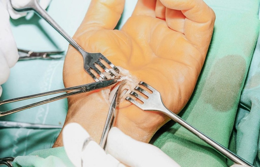
Median nerve surgery in the carpal tunnel corresponds to all the operations carried out to free the median nerve at the level of the wrist, or to repair it in the event of an injury.
The median nerve
It is a nerve that runs from the root of the arm to the hand, crossing the region of the elbow, forearm and wrist. It ends at the level of the hand, dividing into sensitive branches (which give sensitivity to the fingers) and a motor branch (which makes it possible to contract the muscles at the base of the thumb).
The sensitivity of the hand concerned by the median nerve surgery corresponds to the palm of the hand, to the thumb, to the index, to the middle finger and to the lateral half of the ring finger, as well as the distal half of these fingers to their dorsal surface (in red on the diagram).
Compression of the median nerve
It occurs when the pressure exerted on the nerve somewhere in its path is too great for the nerve to function properly. It can be an acute compression, occurring during a major effort, or a chronic compression, evolving over several months or several years. Aside, dynamic compression, represented by the increase in pressure in the muscular compartments during prolonged efforts, as in certain sportsmen and in certain manual work.
The main areas of median nerve pain are in the arm (Struthers arch), in the elbow and the root of the forearm (pronator teres syndrome) and in the wrist (carpal tunnel syndrome). ). The latter is by far the most common cause of nerve compression in humans.
This is the manifestation of pain in the median nerve at the wrist. The carpal bones, which articulate the hand with the forearm, form an arch whose ceiling is formed by a very strong ligament (the anterior carpal ligament). It is therefore more of a “tunnel” than a “channel” (“carpal tunnel syndrome” from the Anglo-Saxons). Since this channel is closed,
Anatomical cut of the carpal tunnel
This channel contains the 9 flexor tendons of the fingers (1 tendon for the thumb, and 2 tendons for each other finger, in black on the anatomical section) and the median nerve, which is a single trunk at this level. At the exit of the carpal tunnel, the nerve divides into several branches towards the muscles of the thumb and the fingers.
Apart from this typical presentation, there are many cases where not all signs are present. These are atypical syndromes, which are not so rar
Neglected, carpal tunnel syndrome can progress insidiously, with progressive muscle wasting at the base of the thumb and a gradual decrease in finger sensitivity. At the final stage, the hand is almost paralyzed, with a flattened appearance and insensitivity of the first 4 fingers.
Carpal tunnel median nerve damage
The median nerve is located below the palm of the hand. He is therefore particularly exposed and vulnerable, especially in the event of a fall on his hands. A wound at this level, accompanied by tingling sensations in the fingers, must require urgent surgical exploration. Wounds from blunt or sharp objects (knives, glass, metal objects) are also at risk of causing damage to the median nerve.
Diagnostic
Your attending physician may, in view of the signs that you describe to him, suspect this carpal tunnel syndrome. He may prescribe you a biological assessment (a blood test) to check that there are no hormonal or metabolic causes, which can trigger or aggravate such a syndrome.
He will entrust you to a specialized surgeon to confirm the diagnosis. Indeed, certain clinical signs when they are found during the clinical examination of the surgeon, allow to evoke with a certain probability this diagnosis.
Confirmation of the diagnosis requires the performance of an electrical examination of the median nerve, as well as the other nerves of the upper limb. This is the electromyogram. This examination, carried out by a neurologist, makes it possible to confirm the suffering of the nerve, to locate it, and to appreciate its severity.
